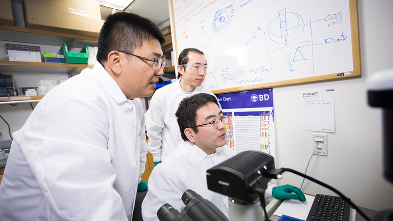
JAX Professor Roel Verhaak (right) and members of his cancer research team at JAX Genomic Medicine in Connecticut. Photo by Cloe Poisson.
1. Finding the cancer dangers that are hidden in plain sight
In the hunt for genetic cancer risk factors, researchers painstakingly analyze huge amounts of genome sequence data. They look for specific mutations that increase or decrease susceptibility, as well as larger-scale genomic patterns that may do the same.
Research has led to tests that identify the mutations driving cancer in individual patients as well as treatments that target those specific cancers.
But all along, there have been cancer‑causing genetic anomalies that remained hidden from standard sequencing protocols. One kind is found within the genome itself: structural variants (SVs), including duplications, insertions, deletions and inversions of normal DNA sequences, that are difficult to spot because they don’t change the sequence data. Recent advances in long‑read sequencing are revealing how common SVs are even in healthy people, and they have also been implicated in multiple cancer types.
Now the significance of another cancer danger is coming to light: extra-chromosomal DNA (ecDNA). This DNA somehow becomes separated from the chromosomes themselves and forms a circular structure in the nucleus. Its segments may contain many copies of various genes, adding to the two copies found, as usual, in the chromosomes themselves. And if ecDNA sequences contain cancer-promoting genes, it can cause havoc.
Cancer initiation
Advanced cancers often have severely disrupted genomes, but the mutation or dysregulation of just a single gene can start cells on the path to malignancy.
A variety of factors can cause cancer-related genes to produce either too much or not enough of a protein. Loss-of-function mutations or deletions of genes such as BRCA1 and BRCA2, which promote the regulation of cell division and DNA repair, and TP53, a tumor suppressor also involved with suppressing cell proliferation, are well-known as cancer risk factors. Other genes, known as oncogenes, become dangerous when they are over-expressed. Oncogenes such as EFGR and Ras produce proteins that promote cell growth, differentiation and proliferation, while myc regulates the expression of many such proliferation-related genes.
When a DNA segment breaks away to form ecDNA, it loses the usual systems that control gene expression. Therefore, ecDNA can be highly transcribed and, if it contains any coding genes, yield an abnormally large amount of protein. When those genes include oncogenes, the normal checks and balances on cell growth and division can be overwhelmed by the uncontrolled protein production, leading to cancer initiation and tumor formation.
Implicated in cancer
It has become increasingly recognized that genomic lesions, which increase oncogene expression, play a role in some cancers, including SVs within chromosomes and ecDNA formation. Researchers first spotted ecDNA in cancer cells decades ago in quite a literal fashion, using fluorescent probes that bound to specific sequences and lit them up, showing that they were physically separate from the typical chromosomal structures in the nucleus. There were still questions surrounding both the prevalence of ecDNA in cancer and the role(s) it might play in disease progression and treatment strategy.
JAX Professor Roel Verhaak has been investigating aspects of ecDNA, particularly in the context of glioblastoma, a brain cancer that is highly resistant to conventional cancer therapies. He found that ecDNA is unevenly inherited by daughter cells during cancer cell divisions, creating important differences between cell populations. It makes sense — while chromosomes are evenly divided in a highly controlled process during cell division, ecDNA has no such regulation and is passed to daughter cells seemingly at random. The differences increase as the cells continue to divide, creating what is known as genomic heterogeneity within the tumor. If there are many sub-populations of cells with different properties, it becomes very difficult to find an effective therapy strategy to eliminate all of them. Therefore, ecDNA not only contributes to glioblastoma initiation, it can also provide a mechanism for therapy resistance and tumor recurrence.
More recently, Verhaak and colleagues took a broader view to assess ecDNA frequency and clinical impact across multiple cancer types in thousands of patients. They found that ecDNA is far more common in a variety of cancers than previously thought — by more than 15-fold— and while its presence is often amplified in cancer cells, it is not in normal tissues. Furthermore, patients whose cancers carry ecDNA with increased oncogene expression have more aggressive cancers than those without. Indeed, ecDNA amplification in tumors was associated with significantly worse five-year survival outcomes.
Is ecDNA a key to stopping therapy-resistant cancers?
The discoveries are a crucial first step toward finding preventions or effective therapies for these dangerous cancers. The insight into ecDNA’s prevalence and importance will fuel exploration into potential clinical targets. For example, how does ecDNA form? Can it somehow be neutralized before it can cause harm? Also, how can diagnosing ecDNA-positive cancers help guide treatment strategies? The findings that ecDNA may underlie some of the most difficult-to-treat cancers indicate that new therapeutic approaches are needed to improve patient prognoses.
While learning that ecDNA increases cancer lethality may be alarming, knowledge is power.
“We are on a mission to improve the outcomes of patients with cancer,” says Verhaak, “and our ecDNA discoveries are pivotal for achieving those goals.”
 JAX scientist Gary Ren (left) and colleagues at his Maine-based cancer research laboratory. JAX photo by Tiffany Laufer.
2. Derailing cancer metastasis
'Metastatic cancer is the most dangerous and remains the most lethal.'
As cancer therapies improve and grow ever more precise, many cancers can be eradicated or effectively shut down at their site of origin. While medically serious, these primary tumors usually have growth pathways that are targetable, and there are now many effective treatments available. Nonetheless, there were 9.5 million cancer deaths worldwide in 2018, and that number is expected to jump to 16.4 million by 2040. So why do so many patients still die?
Unfortunately, not all cancer cells are the same, even within the tumor, and not all of them stay put. Some break away from the primary tumor and move to other locations in the body, in a process known as metastasis. Metastatic cancer is the most dangerous and remains the most lethal. Understanding the processes and variables underlying metastatic cancer and how it might be stopped is therefore essential if we are to make the next leap forward in improving cancer care.
How does cancer spread?
When cancer begins, it usually involves a single rogue cell. Somehow the brakes come off the carefully controlled cell division cycle, allowing the cell, and eventually its many descendants, to grow and divide with relentless speed. As it grows, it quickly adapts to and co-opts biological systems within its particular organ or tissue.
At a certain point, however, cancer cells begin to move, either by growing into other tissues or separating from the original tumor. Cells that break away travel through the blood stream and lymph system to other places in the body. These cells face long odds, and most of them are eliminated by the immune system or fail to adapt to the environment in which they settle. Sometimes they escape immune surveillance for long enough, and adapt to their new environment quickly enough, to survive the transition. Eventually—weeks, months or even years later—they begin to thrive once again and grow aggressively, resistant to treatments that may have worked well for the primary tumor.
Cancer researchers are investigating the steps in the metastatic process to look for ways to stop cancer from spreading. Could immune surveillance be enhanced to eliminate cells before they reach other areas of the body? Could the environments in other tissues be made less hospitable to the cells that infiltrate them? And if they’re not eliminated before reaching a destination, is there a way to maintain metastatic cells in a senescent state, keeping them inactive by undermining their ability to grow and divide? If the answer is yes, and a therapy can be developed that prevents metastatic cancer growth, it would save many lives.
Immune cell roles
Assistant Professor Guangwen “Gary” Ren investigates cancer micro-environments, the normal tissues and cells immediately adjacent to cancer cells and tumors. There are many interactions between cancer cells and their immediate cellular neighbors, some of which are quite important for cancer growth.It’s particularly imperative for the cancer cells to evade and eventually co-opt the local immune cells that might otherwise recognize and destroy them. Not many accomplish this feat, but those that do are able to grow while actually being protected by the immune cells that would normally kill them.
When metastatic cancer cells migrate elsewhere in the body, they’re on their own, and have to deal with a new environment and immune cells. So how are they able to succeed in there, so to speak? Ren is investigating the complex interplay between immune cells and cancer cells, and between different kinds of immune cells, that can dictate success or failure of metastatic cancer spread. Of particular interest are cells known as neutrophils, which are part of the innate immune system, the first line of defense against invading pathogens. Recent research in Ren’s laboratory has shown that neutrophils also play surprising roles in cancer metastasis.
Neutrophil paradoxes
Ren and his team worked with specialized mouse models to look at breast cancer cells that migrate to lung tissue, a common site for breast cancer metastatic spread. They are often eliminated by either neutrophils or natural killer (NK) cells, another component of the innate immune system. In an unexpected result, however, Ren found that when both neutrophils and NK cells are present in lung tissue, the neutrophils don’t react against the cancer cells. Instead, they actually inhibit NK activity, helping the metastatic cells survive and increasing the chance of cancer spread. The finding helps to explain why treatments that increase neutrophil counts in cancer patients, which are often greatly reduced by chemotherapies, are associated with higher risk for subsequent metastatic disease.
In addition, the research team discovered that neutrophils in the lung tend to stock up on fuel in the form of lipids when a breast cancer tumor is growing. Interestingly, the process is stimulated by molecular signals sent from the tumor itself, even before its cells begin to migrate elsewhere. Then, when circulating cancer cells do arrive in the lung, the neutrophils transfer the lipid fuel to them, increasing their ability to survive and proliferate.
Targets for treatment
More research is needed into exactly why neutrophils function as both safeguards against metastasis and, in different contexts, as part of the support system for it. Nonetheless, Ren’s findings provide intriguing targets for therapy refinement and development. For example, assessing NK/neutrophil populations can help inform whether or not to increase neutrophil counts following initial therapy. And disrupting the signaling/metabolic cascade that helps fuel metastatic cancer cells in the lung could greatly reduce the chances for successful spread. These and other treatment regimens are both parts of the larger effort to derail cancer spread and, ultimately, deaths from metastatic cancer.
 John Pierce is a cancer patient who is participating in the Maine Cancer Genomics Initiative. Pierce was able to take part in the MCGI tumor board meeting online in March. Photo by Alexandra Giardino.
3. Creating a model for personalized cancer care
From cancer genomic testing to clinical trials, John Pierce is exploring the latest advances to address his medical issues.
What advice would John Pierce give to someone who has just received a cancer diagnosis?
"First, be your own best advocate. When I was a 20-year-old combat helicopter pilot in Vietnam, I learned that when someone or something is trying to kill you every day, you recognize that nothing focuses your mind like your own mortality."
Second, he says, "get genomic profiling."
Nearly 50 years after his combat experience, Pierce is now retired from a successful and varied career as an internet consultant. And following a series of unusual health crises that trained him to seek out the best medical advice and treatment, he has been diagnosed with cholangiocarcinoma — a very rare cancer of the bile ducts, the slender tubes that carry the digestive fluid bile through the liver. Pierce is now bringing his lifelong facility for quickly acquiring technical expertise to his treatment regime, to be his own best advocate.
Pierce may not be your average cancer patient, but every patient, and every cancer, is genomically unique. Pierce says that when he learned he had cancer, “my first inclination was to obtain genomic testing. Cancer isn’t liver cancer or lung cancer; it’s defined by whatever the cancer’s genomic profile is. And that’s why I started down this path and requested testing.”
maine-cancer-genomics-initiative
Pierce’s oncologist is Dr. Roger Inhorn of MaineHealth, a steering committee member of the Maine Cancer Genomics Initiative (MCGI). JAX founded MCGI in 2016 with a grant from the Harold Alfond™ Foundation and has enrolled every oncology practice in Maine and most of its oncologists. In phase one of the program, which concluded at the end of 2020, participating oncologists submitted patient tumor samples to be sequenced and profiled for genes known to be associated with various cancers, and with response or resistance to approved targeted therapies or new drugs in development approved by the U.S. Food and Drug Administration.
“For almost all of my patients who have an advanced malignancy and have enough tissue available,” Inhorn says, “I offered participation in MCGI. Genomic profile testing helps clarify potential options by looking for targetable mutations for which there might be either a clinical trial option or a commercially available drug that can be used to treat their malignancy.”
Inhorn notes that like many patients participating in MCGI, Pierce “understands that there is also an altruistic piece to this. They understand that MCGI and the clinical community are trying to learn and create a larger database of treatment options and to engage and educate oncologists about how to best use these platforms. I think the day is coming where everyone who has an advanced malignancy will be offered genomic profiling — it’s going to become a standard of care.”
Tumor profiles from MCGI testing, and their best treatment options, are reviewed at Genomic Tumor Board (GTB) sessions, virtual conferences that link clinicians with experts in cancer genomics and clinical trials. Inhorn mentioned to Pierce that there was a tumor board meeting coming up, and Pierce said, “I’d like to participate. I’d like to be in the room when they’re discussing my case.”
Dr. Jens Rueter, chief medical officer at JAX, says that Pierce’s participation in the tumor board “worked out really well. Beforehand I was a little bit nervous about it because I didn’t really know what to expect. He’s an unusual individual because he embraces new technologies, and that’s coupled with really wanting to impact his treatment plan dramatically.”
Rueter says GTBs are an important component of MCGI, as they often provide the most significant input to clinicians with respect to applying the genomic information in their patients’ care plan. Typically, four cases are discussed during each one‑hour meeting. A brief case presentation by the treating oncologist is followed by a presentation of the genomic case information. Then, external advisors specializing in precision oncology give their interpretation of the case and provide significant guidance to the oncologist and the medical team. In this case Pierce himself was also able to query the experts.
Genomic testing identified two targetable mutations in Pierce’s cancer. He is now on his second targeted therapy (erlotinib) after four months on the IDH2 inhibitor enasidenib.
According to Rueter, most cancer patients fall into one of two categories. “The first are comfortable doing what their doctor says, maybe asking questions or even questioning some decisions, but basically trusting the doctor. And the second tend to shop around for doctors until they find the one who tells them what they want to hear.” Pierce is in a rare third category, Rueter says. “He just wants to know everything that could possibly be done to address his cancer.”
Pierce is continuing to actively advocate for his health. “I think cancer and oncology clinicians today don’t talk about cures; they talk about control. And as far as I’m concerned, control isn’t really good enough for what I’m looking for. I am looking for a cure.”
A model for personalized cancer care in a rural setting
MCGI is a statewide collaboration of JAX scientists and community oncologists that brings innovative cancer genomic testing, education and drug access infrastructure to Maine. Every oncology practice in the state is a partner in the program. Initially driven by the need for greater availability of cancer genomic testing in Maine, MCGI has become a model for community precision oncology, or personalized cancer care, in a rural setting. Precision oncology uses analysis of a patient’s normal genetics and the specific mutations found in his or her tumors to guide more targeted treatments.
“Over the last four years, we’ve made great progress with precision medicine in Maine, especially in rural areas of the state,” said Rueter. “In the first phase of the initiative, we provided genomic tests to over 1,600 cancer patients, affecting patient lives from Caribou to Kittery. Over the next five years, we will focus our efforts on helping patients navigate the steps of entering genome-informed clinical trials and of accessing targeted therapies as part of their routine medical care.”
The initiative also plans to expand its reach to other areas of the northeast United States beyond Maine.